Abstract
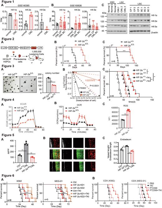
Although the advent of tyrosine kinase inhibitors (TKI) drugs significantly increased the long-term survival of chronic myeloid leukemia (CML), residual LSCs can still lead to drug resistance and disease recurrence. As LSCs in CML rely on hypoxia-inducible factors (HIF) to maintain their own energy metabolism in the hypoxic environment of bone marrow, HIF-2α has not been systematically explored in CML. Here, we explored how HIF-2α participated in drug resistance and provided a potential new clinical strategy for CML. We first investigated the expression of HIF in the database and found the overexpression of HIF-2α in the bone marrow of CML patients compared with normal subjects, while there was no difference in HIF-1α and HIF-3α (Fig. 1A-B). We then collected bone marrow samples from normal subjects and CML patients. We found that compared with normal hematopoietic stem cells (Lin-CD90+CD34+CD38-), CML LSCs (Lin-CD90+CD34+CD38-) had higher levels of HIF-related proteins than common leukemia cells (Fig. 1C).
To further explore the role of HIF-2α in the pathogenesis of CML, we constructed HIF-2α conditional knockout mice through transfecting the plasmid of Cre-BCR-ABL-GFP with transposase and Ctrl-BCR-ABL-GFP without transposition to HSPCs (Fig. 2A). After the first round of leukemia transplantation, the onset of CML was delayed in the HIF-2α knockout mice and the median survival time was prolonged from 31d in the Ctrl group to 60d (Fig. 2B). Next, we harvested fully onset CML mice for the secondary transplantation and found the median survival time extended from 46d to 117d in the HIF-2α knockout group (Fig. 2C).
Then we investigate the biological function of HIF-2α in CML LSCs. We performed the clone formation assay on bone marrow cells of the first round of onset CML mice and found that HIF-2α knockout reduced the colony-forming ability of CML cells (Fig. 3A). To explore the tumorigenic ability of HIF-2α knockout, limiting dilution assay (LDA) was performed and found that the LSCs ratio of CML decreased by 30 times after HIF-2α knockout, from 1/76960 to 1/2237970 (Figure. 3B). Meanwhile, we explored the effect of HIF-2α knockout on the chemosensitivity in CML. TKI treatment combined with HIF-2α knockout can significantly improve the efficacy of TKI and extend the mouse survival time (Figure. 3C). To explore the specific mechanism by which HIF2A affects LSCs, we firstly investigate the effect of HIF-2α on LSCs metabolism. We detected metabolism-related indexes of LSCs in the CML model and found that the glycolysis ability of LSCs was weakened after HIF-2α knockout (Fig. 4A). At the same time, its oxidative phosphorylation pathway and reactive oxygen species (ROS) were enhanced (Fig. 4B-C) which may be an important cause of LSCs apoptosis.
As HIF-2α can directly promote the transcription of pro-angiogenic factors, we detected the content of vascular endothelial growth factor (VEGF) α in the supernatant of mice bone marrow and found that it was significantly increased after the onset of CML but decreased after HIF-2α knockout (Fig. 5A). Fluorescent staining of bone marrow sections revealed that the density of blood vessels in bone marrow increased, whether in the intima (Fig. 5B) after the onset of CML. Next, we treated CML mice with VEGF Monoclonal antibody and found that it alone could prolong the survival time. However, the VEGF Monoclonal antibody did not affect HIF-2α knockout CML mice (Fig. 5C). These results suggest that HIF-2α is involved in bone marrow vascular remodelling after the onset of CML.
To investigate the functional effect of HIF-2α in human CML, the CDX CML mouse model was made with CML cell lines (K562 and MEG-01) to observe the tumorigenesis of CML in nude mice, and they were treated with TKI. We found that HIF-2α knockout could delay the onset of CML and improve the therapeutic effect of TKI (Fig. 6A). To better apply the effect of HIF-2α on CML chemosensitivity to the clinic, we explored the effect of inhibitors of HIF, PT2399, on CML chemosensitivity. PT2399 can significantly increase the chemosensitivity of CML cell lines to TKIs and prolong the survival curve of CML mice treated with TKIs (Fig. 6B).
In conclusion, HIF-2α knockout could inhibit the function of LSCs by affecting their metabolic pattern and vascular microenvironment in CML. Therefore, targeting HIF-2α may become a new strategy, and its specific inhibitor PT2399 may be a candidate drug for clinical application in CML treatment.
Disclosures
No relevant conflicts of interest to declare.
Author notes
Asterisk with author names denotes non-ASH members.


This feature is available to Subscribers Only
Sign In or Create an Account Close Modal